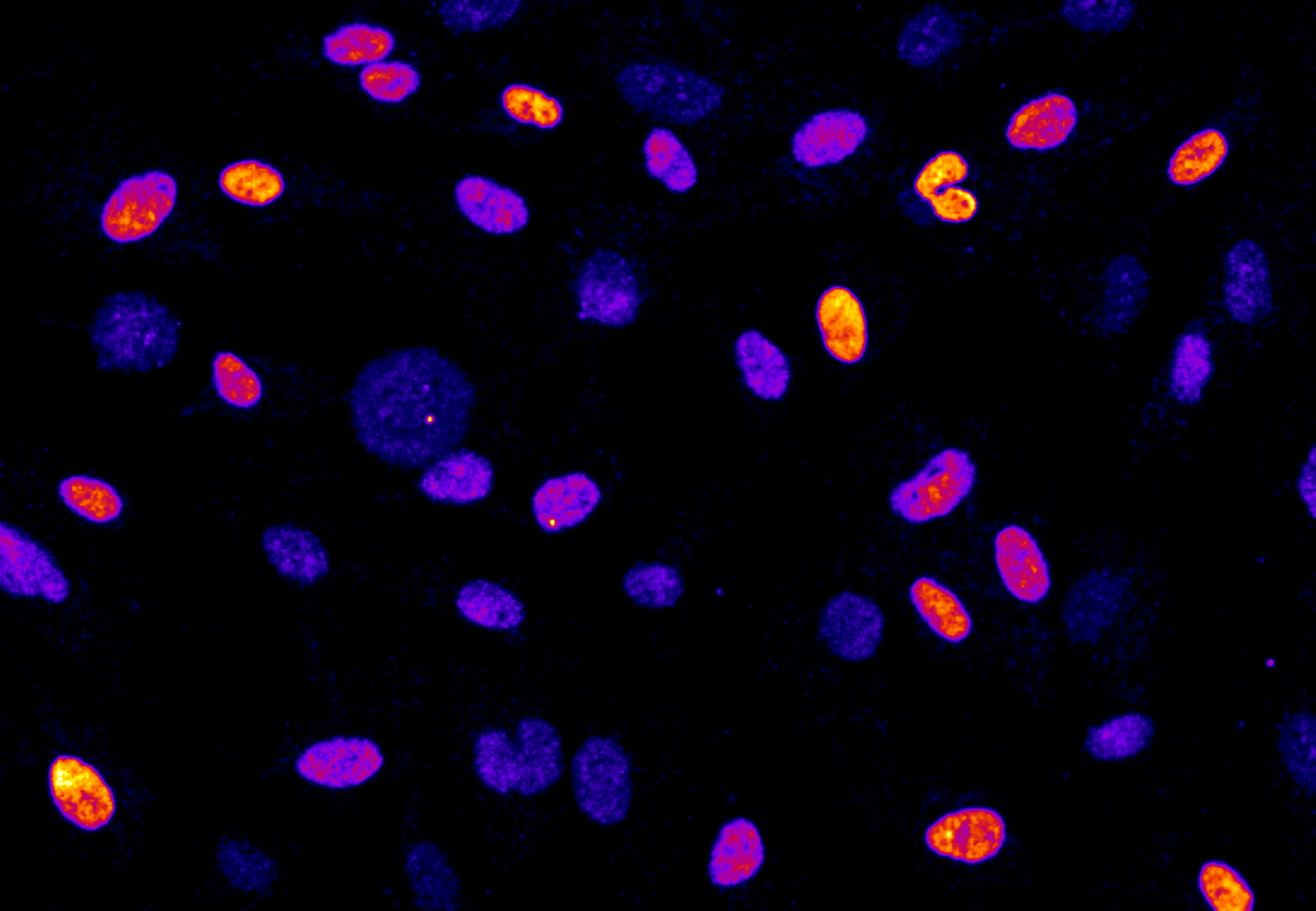
The cells in this microscopy image have been stained with a fluorescent dye that binds only to proteins expressed in defective cells. This technique a...
This microscopy image shows living cells at different stages of the cell cycle. Cells with distinct green spots are currently replicating their DNA, w...
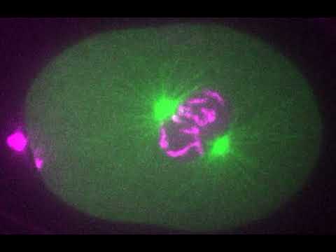
This video captured the first division of a C. elegans worm embryo. The microtubules (green) pull the chromosomes (magenta) toward both poles, centros...