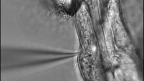
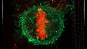
During cell division, the genetic material inside an animal cell assembles along the cell equator. In order for a cell to continue dividing after this...
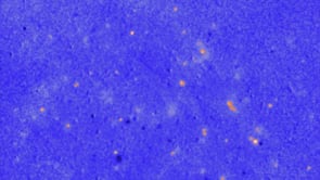
OIST scientists have gained new insight into the abnormal brain activity underlying Parkinson’s disease. In a mouse model of the disease, the scient...
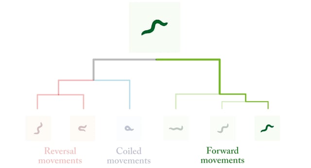
Researchers from the Biological Physics Theory Unit and Vrije Universiteit Amsterdam conducted local linear analyses to reduce the complex posture mov...